


In silico Evaluation of the Active Compounds of Hibiscus sabdariffa Linn as IL-11 Inhibitor
Article Sidebar
Views | PDF/EPUB Downloads:
305
/ 203
/ 71
Main Article Content
Abstract
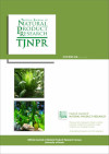
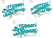
Interleukin (IL) 11 is a pro-inflammatory and pro-fibrotic cytokine. Recently, it has been known that IL-11 inhibition can increase health and lifespan. There are still limited compounds that become IL-11 inhibitors. Hibiscus sabdariffa Linn. (HSL) is a common herbal drink in Asia with good antioxidant and anti-inflammatory properties. This in silico study aims to determine the candidate bioactive compounds in HSL that can act as IL-11 inhibitors. The Protein Data Bank (PDB) database with code 6O4O provided the IL-11 protein, while the PubChem database provided the active compound of HSL. The molecular docking results were displayed using PyMol version 1.3. The best candidates were assessed based on affinity prediction, ADMET profiles, and Lipinski’s rules of five criteria. Quercetin 3-7-diglucuronide, delphinidin 3-O-beta-D-sambubioside, and quercetin 3-rutinoside have shown strong interactions with the targeted protein IL-11 with the least docking score (-7.4~-7.0 kcal/mol). All those phytochemicals interacted with several active sites of IL-11. Quercetin 3-7-diglucuronide interacts with the amino acids Arg33, Arg40, Gly47, Asp48, His49, His161, Trp166, and Arg169. Delphinidin 3-O-beta-D-sambubioside interacts with the amino acids Arg40, Asp48, Gly158, His161, and Arg169. Quercetin 3-rutinoside interacts with the amino acids Arg40, His49, His154, His161, Asp165, and Arg169. Although drug-likeness only met one of five of Lipinski’s rules, ADMET profiles are promising and can be further investigated as possible IL-11 inhibitors within in vitro and in vivo studies to prolong the health and lifespan of mammals.
Downloads
Article Details
Section

This work is licensed under a Creative Commons Attribution-NonCommercial-NoDerivatives 4.0 International License.
How to Cite
References
1. Tenchov R, Sasso JM, Wang X, Zhou QA. Antiaging strategies and remedies: A landscape of research progress and promise. ACS Chem Neurosci. 2024;15(3): 408-446. Doi: doi: 10.1021/acschemneuro.3c00532. DOI: https://doi.org/10.1021/acschemneuro.3c00532
2. Melzer D, Pilling LC, Ferrucci L. The genetics of human ageing. Nat Rev Genet. 2020;21(2):88–101. Doi: 10.1038/s41576-019-0183-6. DOI: https://doi.org/10.1038/s41576-019-0183-6
3. Singh D. Astrocytic and microglial cells as the modulators of neuroinflammation in Alzheimer’s disease. J Neuroinflammation. 2022;19(1):206. Doi: 10.1186/s12974-022-02565-0. DOI: https://doi.org/10.1186/s12974-022-02565-0
4. Birch J, Gil J. Senescence and the SASP: many therapeutic avenues. Genes Dev. 2020;34(23–24):1565–1576. Doi: 10.1101/gad.343129.120. DOI: https://doi.org/10.1101/gad.343129.120
5. Li X, Li C, Zhang W, Wang Y, Qian P, Huang H. Inflammation and aging: signaling pathways and intervention therapies. Signal Transduct Target Ther. 2023;8(1):239. Doi: 10.1038/s41392-023-01502-8. DOI: https://doi.org/10.1038/s41392-023-01502-8
6. Metcalfe RD, Aizel K, Zlatic CO, Nguyen PM, Morton CJ, Lio DSS, Cheng H, Dobson R, Parker M, Gooley P, Putoczki T, Griffin M. The structure of the extracellular domains of human interleukin 11α receptor reveals mechanisms of cytokine engagement. J Biol Chem. 2020;295(24):8285–8301. Doi: 10.1074/jbc.RA119.012351. DOI: https://doi.org/10.1074/jbc.RA119.012351
7. Widjaja AA, Lim WW, Viswanathan S, Chothani S, Corden B, Dasan CM, Goh J, Lim R, Singh B, Tan J, Pua C, Lim S, Adami E, Schafer S, George B, Sweeney M, Xie C, Tripathi M, Sims N, Hübner N, Petretto E, Withers D, Ho L, Gil J, Carling D, Cook S. Inhibition of IL-11 signalling extends mammalian healthspan and lifespan. Nature. 2024;632(8023):157–165. Doi: 10.1038/s41586-024-07701-9. DOI: https://doi.org/10.1038/s41586-024-07701-9
8. Carrillo-Martinez EJ, Flores-Hernández FY, Salazar-Montes AM, Nario-Chaidez HF, Hernández-Ortega LD. Quercetin, a flavonoid with great pharmacological capacity. Molecules. 2024;29(5):1000. Doi: 10.3390/molecules29051000. DOI: https://doi.org/10.3390/molecules29051000
9. Riaz G, Chopra R. A review on phytochemistry and therapeutic uses of Hibiscus sabdariffa L. Biomed Pharmacother. 2018;102:575–586. Doi: 10.1016/j.biopha.2018.03.023. DOI: https://doi.org/10.1016/j.biopha.2018.03.023
10. Bayani GF El, Marpaung NLE, Simorangkir DAS, Sianipar IR, Ibrahim N, Kartinah NT, Mansur I, Purba J, Ilyas E. Anti-inflammatory effects of Hibiscus sabdariffa Linn. on the IL-1β/IL-1ra ratio in plasma and hippocampus of overtrained rats and correlation with spatial memory. Kobe J Med Sci. 2018;64(2):E73–E83.
11. Umeoguaju FU, Ephraim-Emmanuel BC, Uba JO, Bekibele GE, Chigozie N, Orisakwe OE. Immunomodulatory and mechanistic considerations of Hibiscus sabdariffa (HS) in dysfunctional immune responses: A systematic review. Front Immunol. 2021;12:550670. Doi: 10.3389/fimmu.2021.550670. DOI: https://doi.org/10.3389/fimmu.2021.550670
12. Fitrianingrum Y, Indarto D, Kusumawati R, Suselo YH. Actinodaphnine and rutacridone as new T-cell protein tyrosine phosphatase inhibitors for drug development of obesity. IOP Conf Ser Mater Sci Eng. 2019;546(6):062007. Doi: 10.1088/1757-899X/546/6/062007. DOI: https://doi.org/10.1088/1757-899X/546/6/062007
13. Herranz-López M, Olivares-Vicente M, Encinar J, Barrajón-Catalán E, Segura-Carretero A, Joven J, Micol V. Multi-targeted molecular effects of Hibiscus sabdariffa polyphenols: An opportunity for a global approach to obesity. Nutrients. 2017;9(8):907. Doi: 10.3390/nu9080907. DOI: https://doi.org/10.3390/nu9080907
14. Goodsell DS, Olson AJ. Automated docking of substrates to proteins by simulated annealing. Proteins: Structure, Function, and Bioinformatics. 1990;8(3):195–202. Doi: 10.1002/prot.340080302. DOI: https://doi.org/10.1002/prot.340080302
15. Dallakyan S, Olson AJ. Small-molecule library screening by docking with PyRx. Methods Mol Biol. 2015;1263:243–250. Doi: 10.1007/978-1-4939-2269-7_19. DOI: https://doi.org/10.1007/978-1-4939-2269-7_19
16. Schrödinger L, DeLano W. PyMOL. [Online]. 2020 [cited 2025 Feb 5]. Available from: https://pymol.org/2/.
17. Meng XY, Zhang HX, Mezei M, Cui M. Molecular docking: A powerful approach for structure-based drug discovery. Curr. Comput.-Aided Drug Des. 2011;7(2):146–157. Doi: 10.2174/157340911795677602. DOI: https://doi.org/10.2174/157340911795677602
18. Liu K, Zha XQ, Li QM, Pan LH, Luo JP. Hydrophobic interaction and hydrogen bonding driving the self-assembling of quinoa protein and flavonoids. Food Hydrocoll. 2021;118:106807. Doi: 10.1016/j.foodhyd.2021.106807. DOI: https://doi.org/10.1016/j.foodhyd.2021.106807
19. Wu D, Chen Q, Chen X, Han F, Chen Z, Wang Y. The blood–brain barrier: structure, regulation, and drug delivery. Signal Transduct Target Ther. 2023;8(1):217. Doi: 10.1038/s41392-023-01481-w. DOI: https://doi.org/10.1038/s41392-023-01481-w
20. Dulsat J, López-Nieto B, Estrada-Tejedor R, Borrell JI. Evaluation of free online ADMET tools for academic or small biotech environments. Molecules. 2023;28(2):776. Doi: 10.3390/molecules28020776. DOI: https://doi.org/10.3390/molecules28020776
21. Vollmannová A, Bojňanská T, Musilová J, Lidiková J, Cifrová M. Quercetin as one of the most abundant represented biological valuable plant components with remarkable chemoprotective effects - A review. Heliyon. 2024;10(12):e33342. Doi: 10.1016/j.heliyon.2024.e33342. DOI: https://doi.org/10.1016/j.heliyon.2024.e33342
22. Aghababaei F, Hadidi M. Recent advances in potential health benefits of quercetin. Pharmaceuticals. 2023;16(7):1020. Doi: 10.3390/ph16071020. DOI: https://doi.org/10.3390/ph16071020
23. Husain A, Chanana H, Khan SA, Dhanalekshmi UM, Ali M, Alghamdi AA, Ahmad A. Chemistry and pharmacological actions of delphinidin, a dietary purple pigment in anthocyanidin and anthocyanin Forms. Front Nutr. 2022;9:746881. Doi: 10.3389/fnut.2022.746881. DOI: https://doi.org/10.3389/fnut.2022.746881
24. Sogo T, Terahara N, Hisanaga A, Kumamoto T, Yamashiro T, Wu S, Sakao K, Hou D. Anti‐inflammatory activity and molecular mechanism of delphinidin 3‐sambubioside, a Hibiscus anthocyanin. BioFactors. 2015;41(1):58–65. Doi: 10.1002/biof.1201. DOI: https://doi.org/10.1002/biof.1201