


Induction of JUN and FOS Expression by Gallic Acid Derivatives in MCF-7 Breast Cancer Cells: An in Silico and RT-PCR Study
Article Sidebar
Views | PDF/EPUB Downloads:
0
/ 0
/ 0
Main Article Content
Abstract
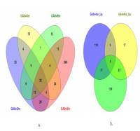
Breast cancer remains the leading malignancy worldwide and continues to cause high morbidity and mortality. Although chemotherapy, hormonal therapy, immunotherapy, and targeted treatments are available, these therapeutic options frequently induce adverse effects and contribute to resistance, limiting long-term outcomes. Gallic acid and its derivatives have been widely reported to exert cytotoxic and pro-apoptotic effects on various cancer cell lines, including MCF7 breast cancer cells. However, the underlying molecular mechanisms, particularly those involving apoptosis-related gene modulation, have not been fully elucidated. This study investigated the expression changes of key apoptosis-related genes in MCF7 cells following treatment with two gallic acid derivatives, N-octyl gallamide and N-tert-butyl gallamide, using integrated in silico and in vitro approaches. Differentially expressed genes (DEGs) were identified from the GSE158788 dataset using GEO2R. Protein–protein interaction networks were constructed using Cytoscape, and KEGG pathway enrichment was performed to determine relevant signaling pathways. JUN and FOS, identified as hub genes, were validated using quantitative RT-PCR in MCF7 cells treated with IC₅₀ and 2×IC₅₀ concentrations of the test compounds. DEG and PPI analyses identified JUN and FOS as key hub genes associated with gallic acid–mediated apoptosis. RT-PCR results demonstrated that N-octyl gallamide significantly upregulated JUN (ΔΔCTs 1.25 ± 0.251; p < 0.05) and FOS (1.82 ± 0.691; p < 0.05), exceeding the effects of tamoxifen. In contrast, N-tert-butyl gallamide did not significantly alter JUN or FOS expression (ΔΔCTs 0.76 ± 0.053; p > 0.05). N-octyl gallamide exhibits strong potential as an anticancer agent through JUN and FOS activation in MCF7 cells. These findings highlight its promise for further development as an alternative or adjuvant breast cancer therapy.
Downloads
Article Details
Section

This work is licensed under a Creative Commons Attribution-NonCommercial-NoDerivatives 4.0 International License.
How to Cite
References
1.GLOBOCAN. The Global Cancer Observatory - Breast cancer fact sheet in Indonesia. International Agency for Research on Cancer - WHO. 2020.
2.GLOBOCAN. The Global Cancer Observatory - Breast cancers fact sheet in the world. International Agency for Research on Cancer - WHO. 2020 [cited 2022 Mar 20]. Available from: https://gco.iarc.fr/today/home
3.Xiao YF, Jie MM, Li BS, Hu CJ, Xie R, Tang B, Yang SM. Peptide-based treatment: A promising cancer therapy. J Immunol Res. 2015;2015:761820–761832.
4.Cui W, Aouidate A, Wang S, Yu Q, Li Y, Yuan S. Discovering anti-cancer drugs via computational methods. Front Pharmacol. 2020;11:1–14.
5.Amayreh M, Fraihat S, Hourani W, Hourani MK. Voltammetric determination of gallic acid and its content in tea samples using modified iodine-coated platinum electrode. Trop J Nat Prod Res. 2021;5(6):1072–1077.
6.Locatelli C, Filippin-Monteiro FB, Creczynski-Pasa TB. Alkyl esters of gallic acid as anticancer agents: A review. Eur J Med Chem. 2013;60:233–239.
7.Tsai CL, Chiu YM, Ho TY, Hsieh CT, Shieh DC, Lee YJ, Tsay GJ, Wu YY. Gallic acid induces apoptosis in human gastric adenocarcinoma cells. Anticancer Res. 2018;38(4):2057–2067.
8.Savi LA, Leal PC, Vieira TO, Rosso R, Nunes RJ, Yunes RA, Creczynski-Pasa TB, Barardi CR, Simões CM. Evaluation of anti-herpetic and antioxidant activities, and cytotoxic and genotoxic effects of synthetic alkyl-esters of gallic acid. Arzneimittelforschung. 2005;55(1):66–75.
9.Arsianti A, Nur Azizah N, Erlina L. Molecular docking, ADMET profiling of gallic acid and its derivatives (N-alkyl gallamide) as apoptosis agent of breast cancer MCF-7 Cells. F1000Res. 2024;11:1453–1478.
10.Yang X, Kui L, Tang M, Li D, Wei K, Chen W, Miao J, Dong Y. High-throughput transcriptome profiling in drug and biomarker discovery. Front Genet. 2020;11:1–12.
11.Chengalvala MV, Chennathukuzhi VM, Johnston DS, Stevis PE, Kopf GS. Gene expression profiling and its practice in drug development. Curr Genomics. 2007;8:262–270.
12.Toro-Domínguez D, Villatoro-García JA, Martorell-Marugán J, Román Montoya Y, Alarcón-Riquelme ME, Carmona-Sáez P. A survey of gene expression meta-analysis: Methods and applications. Brief Bioinform. 2021;22(2):1694–1705.
13.Suryandari DA, Tedjo A, Fadilah S. Differentially expressed genes (DEGs) analysis and in silico studies identify tumor necrosis factor (TNF) inhibition and peroxisome proliferator-activated receptor alpha (PPARA) activation as targets for gallic acid derivatives in insulin resistance. Trop J Nat Prod Res. 2024;8(12):94765–9485
14.Tang HM, Cheung PCK. Gene expression profile analysis of gallic acid induced cell death process. Sci Rep. 2021;11(1):1–17.
15.Tang HM, Cheung PCK. Time-course transcriptomic dataset of gallic acid-induced human cervical carcinoma HeLa cell death. Data. 2025;10(5):61–72.
16.Shannon P, Markiel A, Ozier O, Baliga NS, Wang JT, Ramage D, Amin N, Schwikowski B, Ideker T. Cytoscape: A software environment for integrated models of biomolecular interaction networks. Genome Res. 2003;13(11):2498–2504.
17.Hays A, Wissel M, Colletti K, Soon R, Azadeh M, Smith J, Doddareddy R, Chalfant M, Adamowicz W, Ramaswamy SS, Dholakiya SL, Guelman S, Gullick B, Durham J, Rennier K, Nagilla P, Muruganandham A, Diaz M, Tierney C, John K, Valentine J, Lockman T, Liu HY, Moritz B, Ouedraogo JP, Piche MS, Smet M, Murphy J, Koenig K, Zybura A, Vyhlidal C, Mercier J, Jani N, Kubista M, Birch D, Morse K, Johansson O. Recommendations for method development and validation of qPCR and dPCR assays in support of cell and gene therapy drug development. AAPS J. 2024 Feb 5;26(1):24. doi: 10.1208/s12248-023-00880-9. PMID: 38316745.
18.Jiao Y, Zhao H, Lu L, Zhao X, Wang Y, Zheng B. Transcriptome-wide analysis of the differences between MCF7 cells cultured in DMEM or αMEM. PLoS One. 2024 Mar 28;19(3):e0298262. doi: 10.1371/journal.pone.0298262. PMID: 38547234; PMCID: PMC10977736.
19.Livak KJ, Schmittgen TD. Analysis of relative gene expression data using real-time quantitative PCR and the 2−ΔΔCT method. Methods. 2001;25(4):402–408.
20.Karin M, Liu ZG, Zandi E. AP-1 function and regulation. Curr Opin Cell Biol. 1997;9(2):240–246.
21.Pulverer BJ, Kyriakis JM, Avruch J, Nikolakaki E, Woodgett JR. Phosphorylation of c-Jun mediated by MAP kinases. Nature. 1991;353(6345):670–674.
22.Moodbidri MS, Shirsat NV. Activated JNK brings about accelerated apoptosis of Bcl-2-overexpressing C6 glioma cells on treatment with tamoxifen. J Neurochem. 2005;92:1–9.
23.Mandlekar S, Yu R, Tan TH, Kong AN. Activation of caspase-3 and c-Jun NH2-terminal kinase-1 signaling pathways in tamoxifen-induced apoptosis of human breast cancer cells. Cancer Res. 2000;60:5995–6000.
24.Ahmed NS, Samec M, Liskova A, Kubatka P, Saso L. Tamoxifen and oxidative stress: An overlooked connection. Discov Oncol. 2021;12(1):17-31.
25.Bekele RT, Venkatraman G, Liu RZ, Tang X, Mi S, Benesch MG, Mackey JR, Godbout R, Curtis JM, McMullen TP, Brindley DN. Oxidative stress contributes to the tamoxifen-induced killing of breast cancer cells: Implications for tamoxifen therapy and resistance. Sci Rep. 2016;6:21164–21180.
26.Aborehab NM, Elnagar MR, Waly NE. Gallic acid potentiates the apoptotic effect of paclitaxel and carboplatin via overexpression of Bax and P53 on the MCF-7 human breast cancer cell line. J Biochem Mol Toxicol. 2021;35(2):e22638–e22648.
27.Yeh RD, Chen JC, Lai TY, Yang JS, Yu CS, Chiang JH, Lu CC, Yang ST, Yu CC, Chang SJ, Lin HY, Chung JG. Gallic acid induces G₀/G₁ phase arrest and apoptosis in human leukemia HL-60 cells through inhibiting cyclin D and E, and activating mitochondria-dependent pathway. Anticancer Res. 2011;31(9):2821–2832.
28.Sofi S, Mehraj U, Jan N, Almilaibary A, Ahmad I, Ahmad F, Ahmad Mir M. Clinicopathological significance and expression pattern of Bcl2 in breast cancer: A comprehensive in silico and in vitro study. Saudi J Biol Sci. 2024;31(2):103916-10.